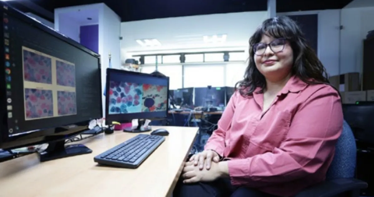

A Mexican researcher, Irari Jiménez López, from the National Polytechnic Institute (IPN), developed an Artificial Intelligence (AI) tool capable of speeding up the detection of cervical cancer through the automated analysis of cytological images.
The researcher, who is also a doctoral student at the Center for Computing Research (CIC), developed a computer program that analyzes images of isolated cells using different color spaces. This strategy makes it possible to reorganize the visual information in an image to highlight characteristics that can facilitate the automatic classification of cells and reduce the influence of less relevant features.
“What we propose is to analyze these cervical cytology images through Machine and Deep Learning models (some of the most advanced areas of AI), as well as computer vision algorithms,” Jiménez López told Xinhua.
Cervical cancer is one of the main public health challenges in Mexico, where it represents the second leading cause of death among women, behind only breast cancer. Therefore, timely diagnosis is crucial to reducing the incidence of the disease.
Model helps classify cellular information
The IPN expert explained that these color models can highlight certain characteristics that can help AI algorithms better classify information from normal and abnormal cells.
“We use an architecture known as a convolutional neural network, which has the ability to analyze images on its own. (…) We use this type of architecture, but again, when it comes to colors, we perform a type of preprocessing,” the university student explained.
She emphasized the importance of this initiative given the challenges that cervical cytology, commonly known as the “Pap smear,” continues to face as one of the main screening methods in the healthcare sector.
The interpretation of samples, the expert continued, requires specialized personnel and can involve long working hours, in addition to possible discrepancies among specialists.
“I believe that something we are developing is analyzing the images as an expert physician would see them; that is, an image with multiple objects. Although there are already studies that analyze cytology images, there are not many studies that use cervical cytology images,” Jiménez López explained.
Technology inspired by human vision: Valdez Rodríguez
For his part, CIC expert José Eduardo Valdez Rodríguez said that the system is developed using AI algorithms, particularly deep neural networks capable of learning from images. Among these are convolutional neural networks, which are inspired by the way human vision works.
These networks analyze images through different layers and progressively identify characteristics, ranging from the most general elements to the finer details.
In this context, the system can learn to recognize patterns in sample images and assist specialists in detecting possible abnormalities.
As a complementary component, the project includes the development of a segmentation algorithm whose purpose is to identify regions of interest within the image in order to delineate cellular structures.
This information makes it possible to perform a quantitative analysis of the image and determine whether it corresponds to a healthy or abnormal cell.
So far, the results obtained show accuracy levels of more than 95 percent in identifying isolated cells, a figure that demonstrates the project’s potential to support the analysis of medical images.

Source: sinemabrgo